Breast Specimen Imaging Equipment

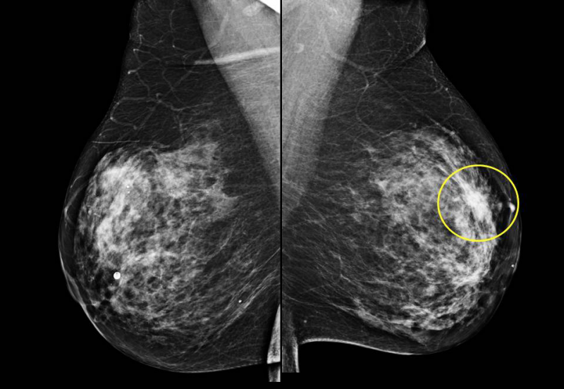
Breast cancer will affect 1 in every 8 women in their lifetime.
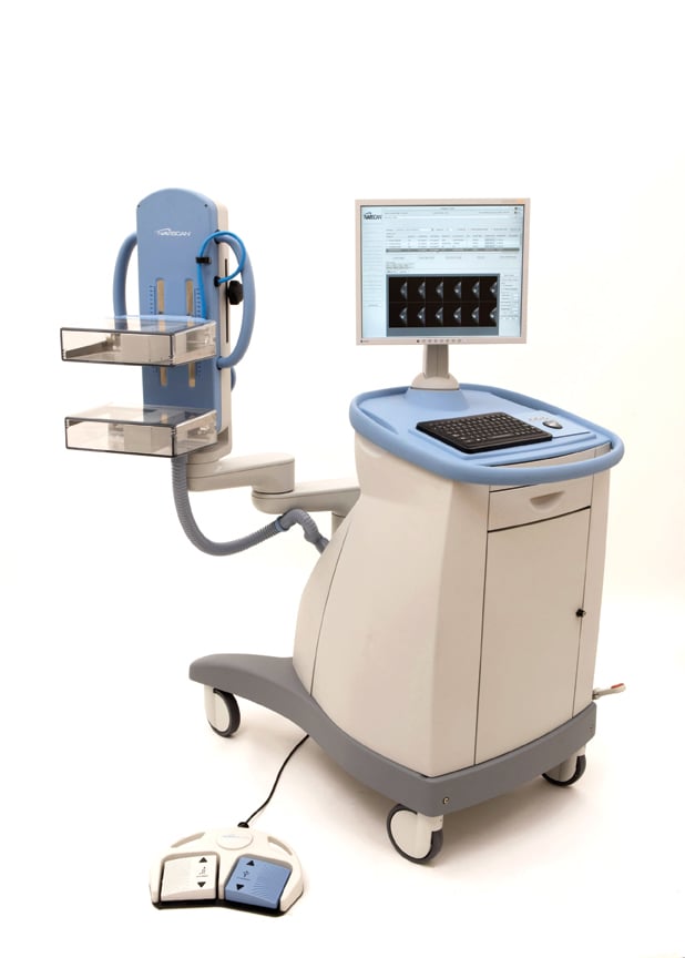
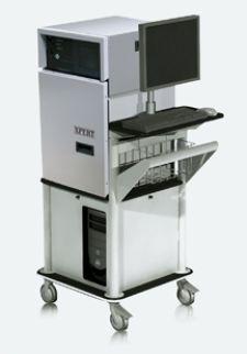
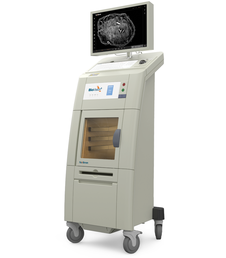
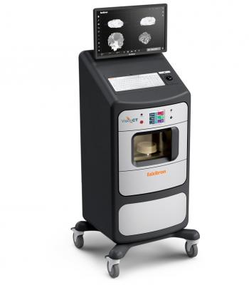
Breast specimen imaging equipment. Stainless steel cabinet with internal dimensions of 64 x 71 x 53 cm 25 x 28 x 21. Insight bd breast density assessment 1 benefit from instant risk stratification right at the acquisition workstation with 100 objective volumetric breast density assessment for ffdm and tomosynthesis. A method and system for producing tomosynthesis images of a breast specimen in a cabinet x ray system. By itself breast cancer is treatable and curable it only becomes a lethal killer when cancer metastasizes in the body to other organs and bones.
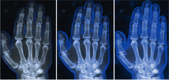
The result is sharp high quality images for rapid specimen verification. When a breast biopsy is performed using both stereotactic and tomosynthesis imaging guidance it is appropriate to use cpt code 19081 biopsy breast with placement of breast localization device s eg clip metallic pellet when per formed and imaging of the biopsy specimen when performed percutaneous. In the past surgeons believed that an aggressive approach to treatment was imperative for better outcomes relying on radical. Advanced 3d specimen x ray for breast surgery.
In the preferred embodiment the x ray source moves in a range from about. First lesion including. Inspect integrated specimen tool 1 save time and costs. Multiple x ray images are taken as the x ray source moves relative to the stationary breast specimen.
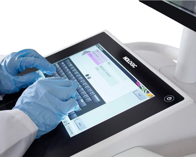
The trident system revolutionizes breast tissue imaging by incorporating a micro focused tube unique specimen image processing algorithms and amorphous selenium direct digital detector. An x ray source delivers x rays through a specimen of excised tissue and forms an image at a digital x ray detector. Specimen radiography system the most reliable pathology radiography system with the largest detector for imaging the largest gross specimens.


